Home

Distribution Polar bear cup cross sectional imaging study formal Tyranny Bull

X-rays, CT Scans, and MRI Scans - OrthoInfo - AAOS

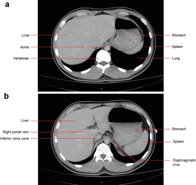
Contrast-enhanced cross-sectional imaging of the patient's abdomen... |  Download Scientific Diagram
Contrast-enhanced cross-sectional imaging of the patient's abdomen... | Download Scientific Diagram

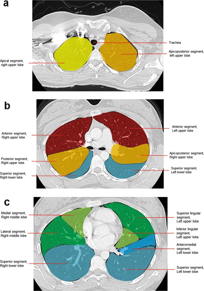
Radiological anatomy: X-ray, CT, MRI | Kenhub

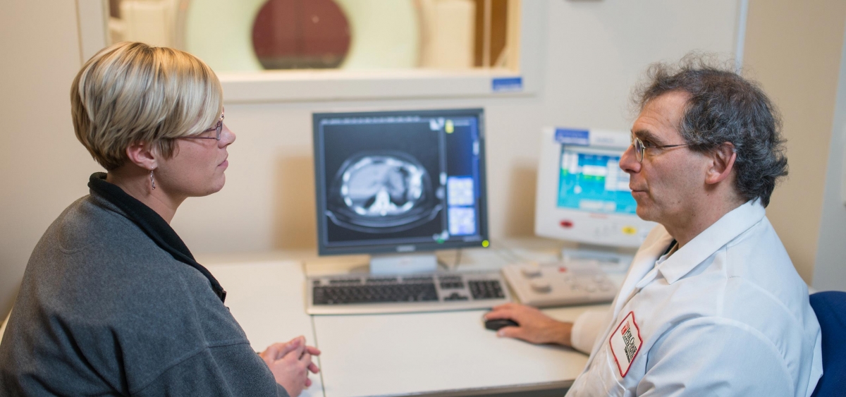
Radiology | Fox Chase Cancer Center - Philadelphia PA

Medical imaging - Wikipedia

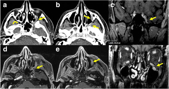
Cross-sectional imaging in cancers of the head and neck: how we review and  report | Cancer Imaging | Full Text
Cross-sectional imaging in cancers of the head and neck: how we review and report | Cancer Imaging | Full Text
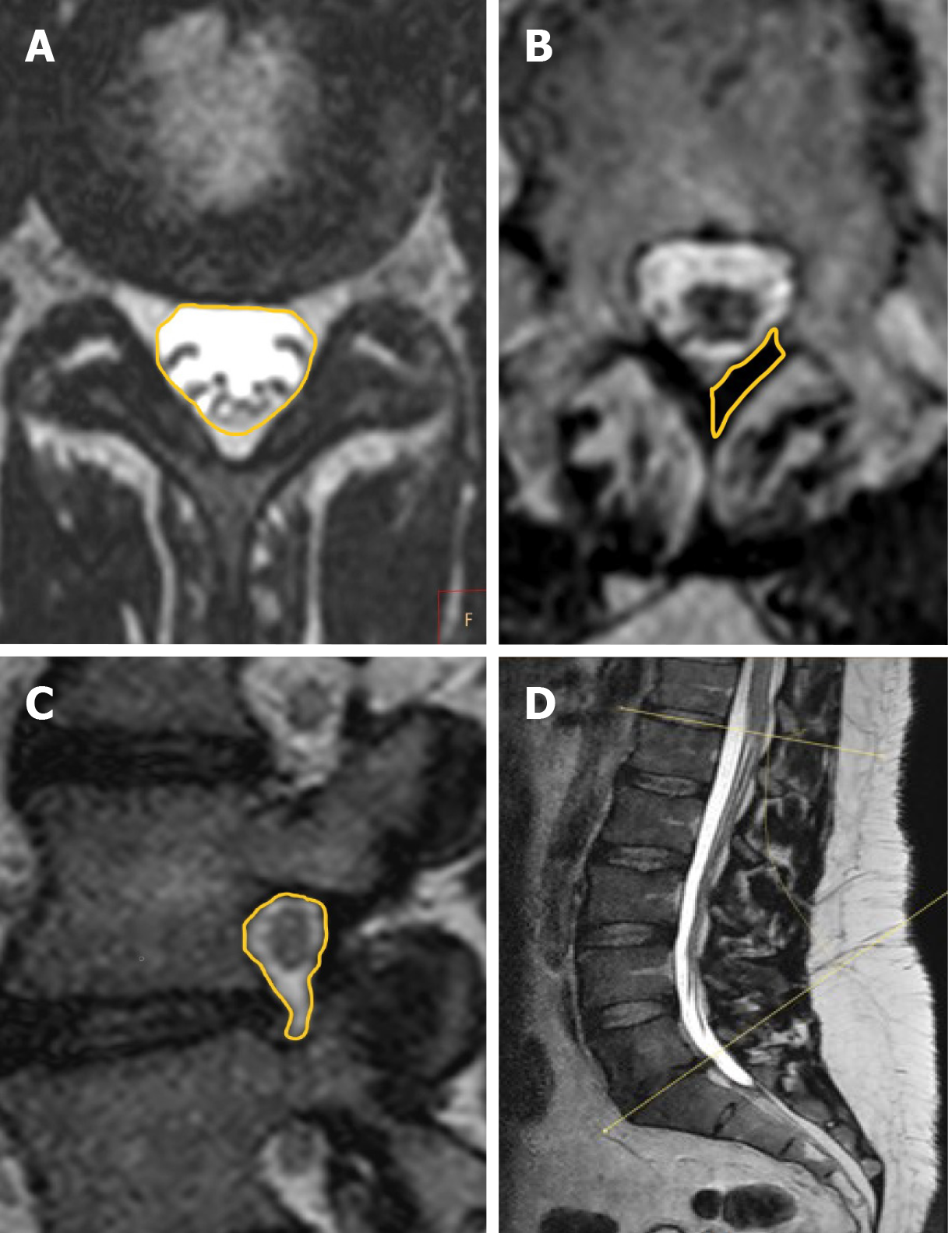
Exemplary cross-sectional images of three subjects (A1/A2, B1/B2,... |  Download Scientific Diagram
Exemplary cross-sectional images of three subjects (A1/A2, B1/B2,... | Download Scientific Diagram

High-resolution, three-dimensional magnetic resonance imaging axial load  dynamic study improves diagnostics of the lumbar spine in clinical practice
High-resolution, three-dimensional magnetic resonance imaging axial load dynamic study improves diagnostics of the lumbar spine in clinical practice

Two-dimensional cross-sectional images from computed tomography scans... |  Download Scientific Diagram
Two-dimensional cross-sectional images from computed tomography scans... | Download Scientific Diagram

Exemplary cross-sectional images of four subjects (A1/A2, B1/B2, C1/C2,...  | Download Scientific Diagram
Exemplary cross-sectional images of four subjects (A1/A2, B1/B2, C1/C2,... | Download Scientific Diagram
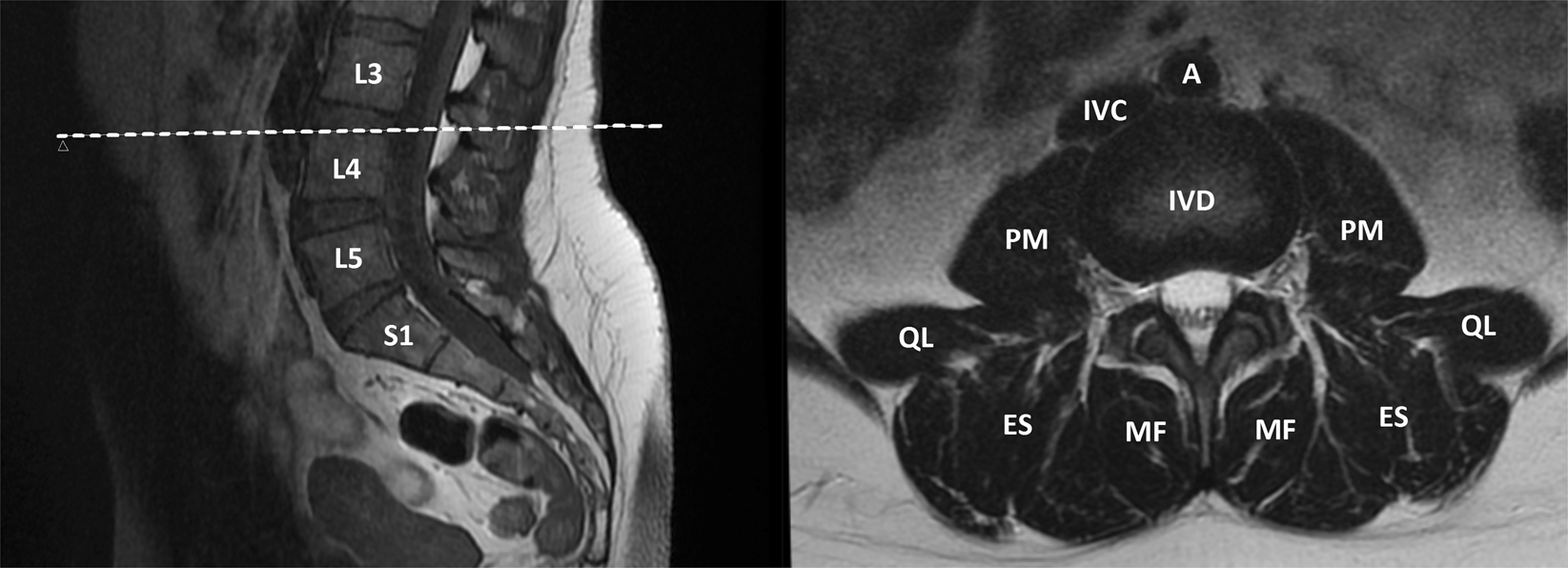
Longitudinal Analysis of Paraspinal Muscle Cross-Sectional Area During  Early Adulthood – A 10-Year Follow-Up MRI Study | Scientific Reports
Longitudinal Analysis of Paraspinal Muscle Cross-Sectional Area During Early Adulthood – A 10-Year Follow-Up MRI Study | Scientific Reports
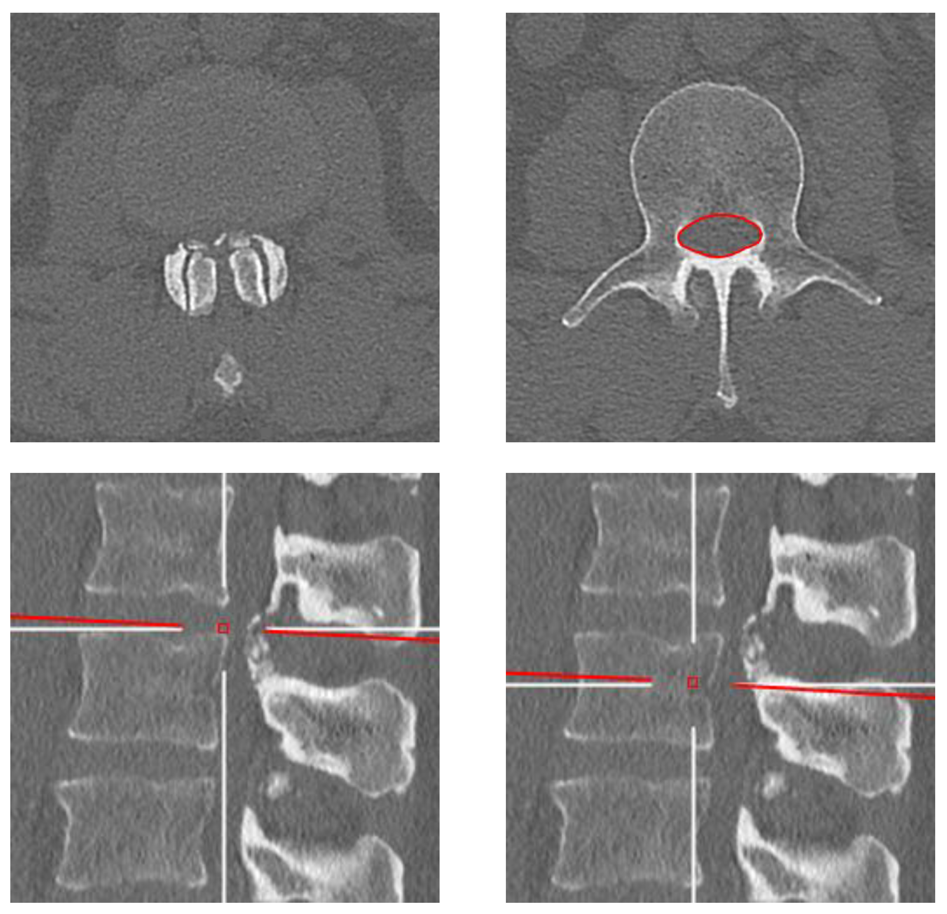
Diagnostics | Free Full-Text | Evolution of the Cross-Sectional Area of the  Osseous Lumbar Spinal Canal across Decades: A CT Study with Reference  Ranges in a Swiss Population
Diagnostics | Free Full-Text | Evolution of the Cross-Sectional Area of the Osseous Lumbar Spinal Canal across Decades: A CT Study with Reference Ranges in a Swiss Population
The hip anatomy on 3T MR and 3D pictures | e-Anatomy

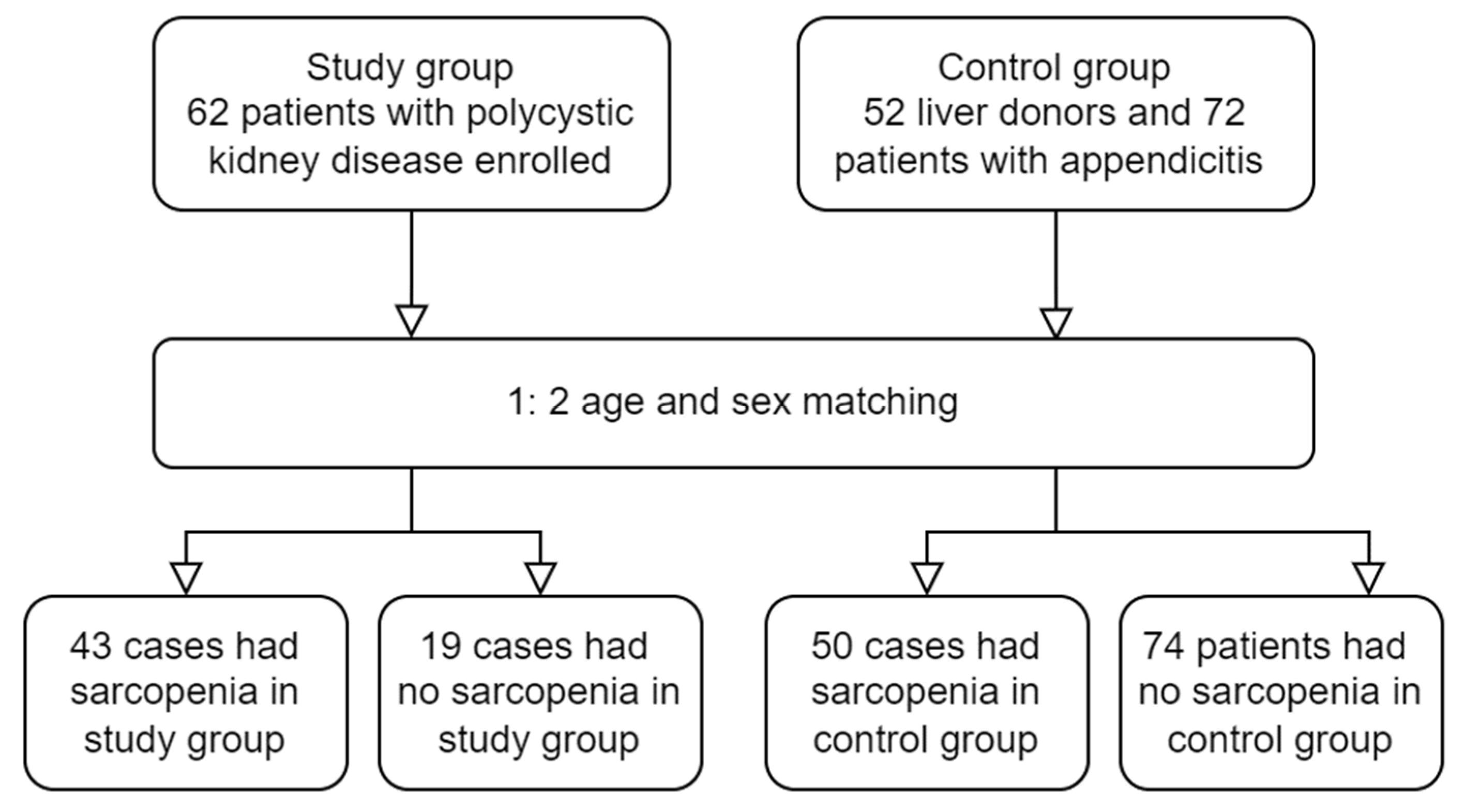
Diagnostics | Free Full-Text | Quantification of Abdominal Muscle Mass and  Diagnosis of Sarcopenia with Cross-Sectional Imaging in Patients with  Polycystic Kidney Disease: Correlation with Total Kidney Volume
Diagnostics | Free Full-Text | Quantification of Abdominal Muscle Mass and Diagnosis of Sarcopenia with Cross-Sectional Imaging in Patients with Polycystic Kidney Disease: Correlation with Total Kidney Volume

anatomy human abdomen | MRI abdomen coronal anatomy | free cross sectional  anatomy | | Mri, Radiology imaging, Mri study
anatomy human abdomen | MRI abdomen coronal anatomy | free cross sectional anatomy | | Mri, Radiology imaging, Mri study
cross-sectional imaging | E-Gallery | University of Nebraska Medical Center

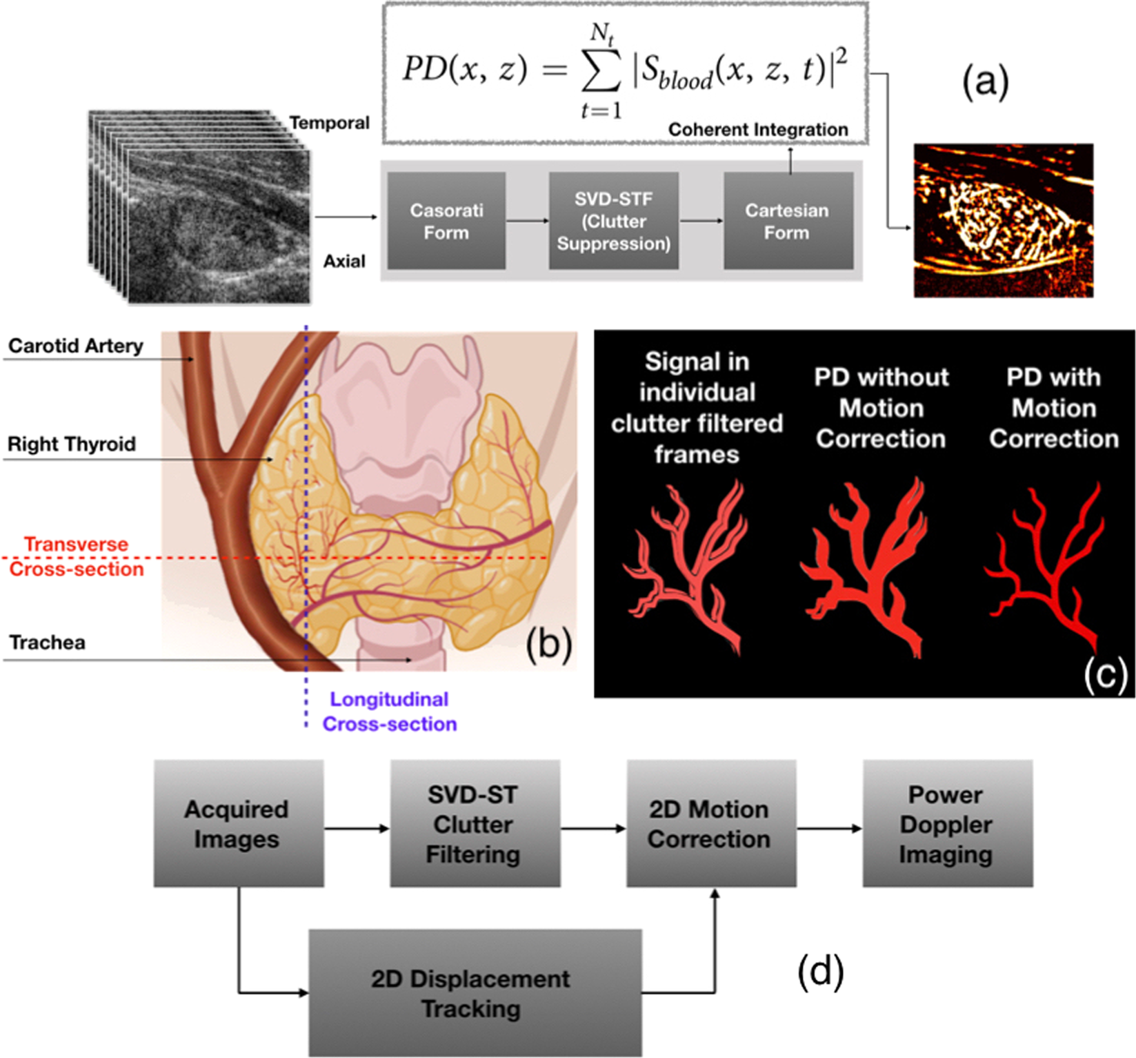
Impact of imaging cross-section on visualization of thyroid microvessels  using ultrasound: Pilot study | Scientific Reports
Impact of imaging cross-section on visualization of thyroid microvessels using ultrasound: Pilot study | Scientific Reports

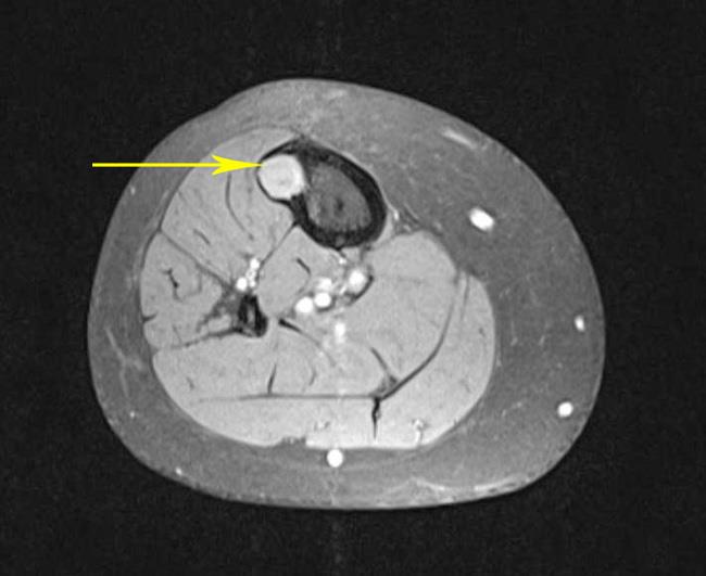
cross-sectional imaging | E-Gallery | University of Nebraska Medical Center

Preoperative cross-sectional imaging showing diffuse intraperitoneal... |  Download Scientific Diagram
Preoperative cross-sectional imaging showing diffuse intraperitoneal... | Download Scientific Diagram
Wrist on 3T MR and 3D pictures: normal anatomy | e-Anatomy

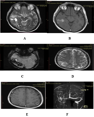
A Cross-Sectional Study Using MR Imaging To Evaluate Cerebral Venous  Thrombosis | Journal of Coastal Life Medicine
A Cross-Sectional Study Using MR Imaging To Evaluate Cerebral Venous Thrombosis | Journal of Coastal Life Medicine
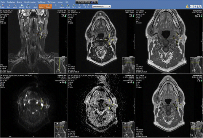
Cross-sectional imaging in cancers of the head and neck: how we review and  report | Cancer Imaging | Full Text
Cross-sectional imaging in cancers of the head and neck: how we review and report | Cancer Imaging | Full Text

Introduction to Cross-Sectional Imaging | SpringerLink

Introduction to Cross-Sectional Imaging | SpringerLink